On this page, you will find a list of the structural biology services available through Instruct-ERIC and associated providers in the ISIDORe project.
To start your application, select the service you would like to access in our catalogue of services and click on the red button “Apply for access” located at the bottom right of your service drop-down.
Find additional information about the ISIDORe transnational call on our ISIDORe help page.
Please contact us if you have any question regarding ISIDORe call and the application process at isidore@instruct-eric.org
Crystallisation allows the 3D structure of macromolecules to be revealed through X-ray diffraction. Instruct offer a fully automated crystallisation pipeline to achieve high-throughput with short crystallisation plate processing times, high reproducibility, and increased efficiency of the screening process.
The 'From Sample to X-ray Diffraction' platform at EMBL Hamburg offers services for obtaining crystal structures from molecules of interest. Services include: sample characterization by various biophyscial methods, sample optimization, high-throughput crystallization, robotic crystal harvesting, diffraction data collection on synchrotron beamlines, data processing, and support in structure solution and refinement.
EMBL Hamburg operates three beamlines at the PETRA III synchrotron ring, two for macromolecular X-ray crystallography and one for small angle X-ray scattering on biological macromolecules. Academic access to the beamlines is available to all research groups, and is prioritised on scientific grounds only. All proposals for beamline access are evaluated by the Project Evaluation Committee.
EMBL beamlines also offer possibilities for mail-in and remote access. Users are encouraged to contact the beamline responsibles for further details.
The Sample Preparation and Characterisation (SPC) facility supports users on the beamline and in addition offers expertise to check and improve the samples that are submitted to the crystallisation facility and the SAXS beamline.
The NKI Protein Facility has a strong record in sample preparation for structural biology, and offers their users access to macromolecular crystallisation:
- Robotics for preparation of nanoliter crystallisation drops in 96-well format.
- Storage of crystallisation plates (4°C or 20°C).
- Screening and ranking of crystallisation conditions in 96-well format (TOPAZ, AutoInspex station).
Solving experimental (crystal, crye-EM) structures of macromolecjules and their complexes is beyond the scope of the NKI Protein facility access services for Instruct-ERIC!
The platform at the Biomolecular Interactions and Crystallography core facility (BIC) at CEITEC Instruct centre provides services for biophysical and structural characterisation of biomolecules, and studying (bio)molecular interactions. It is equipped with the instrumentation to set up crystallisation conditions of biomolecules and their complexes, basic characterisation of physical properties of the molecules (analytical ultracentrifugation, dynamic light scattering, CD spectroscopy, differential scanning calorimetry, differential scanning fluorimetry, size-exclusion chromatography), and to study thermodynamics and/or kinetics of interactions (isothermal titration calorimetry, surface plasmon resonance, bio-layer interferometry, microscale thermophoresis, CD spectroscopy, analytical ultracentrifugation). The facility is accessible each working day upon previous agreement.
More information can be found on the Biomolecular Interactions and Crystallography core facility webpage.
EMBL Grenoble offers access to automated nano-volume crystallization screening and optimization services. Additional services include sample quality assessment and buffer optimization through TSA and estimation of crystallization likelihood. This platform is remotely operated through the web via the Crystallographic Information Management System (CRIMS) providing real-time access to results and experimental parameters.
Biocenter Oulu Protein Crystallography core facility offers crystallization using nanoliter dispensers and a collection of commercial and self maintained crystallization screens. Crystallization is followed using the IceBear software and remote access to monitor the experiments can be provided following the visit.
The high throughput crystallisation platform offers vapour diffusion as well as crystallisation under oil screening and optimisation in sitting drops. Flexible methods have been developed to adapt each step to individual project requirements. At the crystallisation level, we have especially focused on developing techniques to optimise crystal growth.
Crystallization of Proteins and Nucleic Acids core facility provides services focused on obtaining crystals of biomacromolecules and their complexes. We support both manual and robotic crystallization drops setting under various conditions. Remote control of crystal growth in the crystallization hotel is provided using the web interface over Internet.
Expertise
The macromolecular crystallisation platform enables in-drop dynamic light scattering measurement to check the quality of the protein sample, robotic setup of 96-well crystallisation plates, incubation at selected temperature from a wide range, and automated monitoring of the crystallisation experiments. Experiments can be stored at 4-30°C (or higher). Dedicated rooms with stereomicroscopes for crystal manipulation are available at 20°C, 10°C and 25°C (or higher).
Crystallisation screening: Utilizing the sitting and hanging drop by vapor diffusion methods for crystallising soluble proteins, using the Mosquito, and LCP Mosquito crystallisation robots.
The ‘Sample Characterization for Serial Crystallography’ service focuses on evaluating microcrystal suspensions to ensure optimal quality for XFEL data collection. With state-of-the-art equipment and experienced staff, users can characterize their samples in detail and refine preparation strategies accordingly.
This service is to ensure that crystal quality and sample properties are well understood prior to beamtime, thereby improving data collection efficiency and overall experimental success.
Our laboratory is equipped with advanced instruments for comprehensive sample assessment, including Dynamic Light Scattering (DLS) for evaluating particle size distribution, Transmission Electron Microscopy (TEM) for direct visualization of microcrystals and, where feasible, diffraction tests, and Second-Order Nonlinear Imaging of Chiral Crystals (SONICC) for detecting protein crystals in complex mixtures. These techniques enable the detailed analysis of crystal morphology, size, homogeneity, and diffraction potential.
This service can be accessed through both remote and on-site modes, depending on the user’s needs and the nature of the characterization tasks.

This service, Serial Crystallography Sample Preparation, provides expert support for optimal sample preparation using our staff’s expertise and state-of-the-art laboratory equipment.
We provide comprehensive support for the preparation of samples used in serial crystallography experiments. Our expertise covers the entire process from microcrystal growth and optimization, ensuring that samples meet the specific requirements for high-quality serial crystallography experiments.
Users have access to fully equipped laboratory facilities for protein microcrystallization. Facility staff assists researchers in developing and refining crystallization conditions, with particular emphasis on generating stable and uniform microcrystals suitable for serial data collection.
We offer guidance in evaluating crystal quality through microscopic inspection and diffraction testing, and in optimizing parameters such as crystal size distribution and concentration. For users new to the field, tailored consultation and training can be provided to ensure successful sample preparation workflows for XFEL beamtime.
This service is available through both remote and on-site access.

Instruct provides funding to access data analysis tools in addition to tools to collect structural biology data, maximising the output of access visits and heightening the impact of structural biology research.
The Scipion-Chem Service enables researchers to transform structural information about biological macromolecules into actionable hypotheses for ligand binding and therapeutic development. The service provides a fully integrated environment for binding pocket identification, ligand docking, and virtual screening, allowing users to start with experimentally determined or modeled 3D structures and rapidly generate ranked lists of potential ligands. Users can submit their structures and (optionally) their own compound libraries or access curated public collections to explore new binding sites, prioritise hits, and guide experimental validation.
The service requires as input a 3D atomic structure of the target macromolecule. The user may optionally provide a library of small molecules in SMILES format; otherwise, the screening will be performed against a curated set of ~1500 FDA-approved compounds.
Scipion-Chem offers two main types of analyses:
Results are fully integrated within the Scipion environment, providing interactive 3D visualization of druggable sites, binding poses, and ranked compound lists. The outputs can be exported for further refinement, such as molecular dynamics simulations or medicinal chemistry optimization.
This service enables Instruct users to derive early insights into the druggability of newly solved macromolecular structures and to generate experimentally testable hypotheses about potential ligands. The integration into Scipion ensures traceability, reproducibility, and interoperability with other Instruct pipelines, including those for cryo-EM data processing and atomic model refinement.
The Instruct Image Processing Center (I2PC), part of Instruct-ES, offers an extended duration service "FlexibilityHub” targeted to the exploration of the conformational landscape of flexible macromolecules from sets of cryo Electron Microscopy images of purified samples. A continuous flexibility analysis will be performed using recently developed tools (including Zernike3D, developed at I2PC, among several other methods). This service is especially suited for samples presenting large structural flexibility.
This platform will provide personalised support to Instruct projects requiring specialised processing of either electron or X-ray microscopy images. It will also help developers in integrating their methods into generally accessible analysis workflows. The Instruct Image Processing Centre (I2PC) in Madrid will assign personnel to this analysis, as well as the required computational resources.
The high-resolution electron microscope has evolved into a sophisticated instrument that is capable of routinely providing quantitative structural information on the atomic scale.
The facility will provide support and expertise to researchers using different techniques: Cryo-EM sample preparation and characterisation, Cryo-EM image acquisition of unstained biological material for cryo-EM single particle or cryoelectron tomography (cryo-ET), cryo-electron diffraction (microED) and cryocorrelative microscopy (cryo-CLEM). For many of these services, it is advisable to contact the Instruct Image Processing Centre for data processing support).
The Imaging Centre at EMBL Heidelberg gives access to state-of-the-art cryo-EM equipment for structure determination projects using the latest technology and methods in single-particle analysis, cryo-electron tomography (cryo-ET) and Cryo-correlative light and electron microscopy (Cryo-CLEM).
The Instruct Image Processing Center (I2PC), part of Instruct-ES, offers an extended duration service "FlexibilityHub” targeted to the exploration of the conformational landscape of flexible macromolecules from sets of cryo Electron Microscopy images of purified samples. A continuous flexibility analysis will be performed using recently developed tools (including Zernike3D, developed at I2PC, among several other methods). This service is especially suited for samples presenting large structural flexibility.
This platform will provide personalised support to Instruct projects requiring specialised processing of either electron or X-ray microscopy images. It will also help developers in integrating their methods into generally accessible analysis workflows. The Instruct Image Processing Centre (I2PC) in Madrid will assign personnel to this analysis, as well as the required computational resources.
The Cryo-electron microscopy and tomography core facility (CEMCOF) at CEITEC Instruct centre is an open access facility providing access to state-of-the-art instrumentation for cryo-electron microscopy, support with cryo-EM sample preparation, and data analysis. The facility handles BSL1 and BSL2 samples, respectively, and is available to its users for both service and collaborative projects.
More information can be found on the Cryo-electron microscopy and tomography core facility (CEMCOF) webpage
The facility provides access to state-of-the-art cryo-EM instrumentation, including automated workflows for single particle data analysis, electron cryo-tomography, lamella preparation, and correlative imaging.
The cryo-EM facility offers the following workflows:
Tailored support is provided for Instruct users, including project feasibility assessments, proposal preparation, and scheduling support.
The Electron Bio-Imaging Centre (eBIC) provides scientists with state-of-the-art experimental equipment and expertise in the field of cryo-electron microscopy, for both single particle analysis and cryo-tomography. Currently eBIC houses five Titan Krios microscopes, a Talos Arctica, Glacios, and a Scios and Aquilos cryo-FIB/SEM.
The cryoEM facility of the ER-C is situated on the campus of the Forschungszentrum Jülich, a major interdisciplinary research centre and member of the Helmholtz Association. With state-of-the-art equipment, like the Titan Krios G4 (TFS), the facility is specialised in cryoEM sample preparation and imaging of SPA- or cryoET- projects of biological specimen.
The EM platform at IBS-ISBG provides access via Instruct-ERIC to a full range of instruments (three TEMs and auxiliary equipment) to users located at any Instruct member state. This includes our flagship instrument: the Titan Krios G4 microscope CM02; part of the national infrastructure "France cryoEM."
Services include classical negative staining experiments (for quality control or morphological analysis), sample vitrification and sample optimisation for cryo-EM experiments, screening and data acquisition for single particle or tomography experiments.
The PF is also offering cellular EM services for sample preparation (at room or cryo temperatures), sectioning and imaging.
We are currently developing microED and we are interested in collaborations, but this not yet available as a service.
Titan Krios G4 (CM02), available for high-end data acquisition for single particle and tomography.
Talos Glacios, available for cryo-EM sample optimization, screening and for data collection in tomography and single-particle experiments.
Tecnai T12, used for negative staining and cellular electron microscopy.
Tecnai F20, equipped for cryo-EM/ET and microED.
Three Vitrobot MARK IV (including one installed in an anaerobic glove box) and a Leica GP2 for specimen vitrification.
Equipment for freeze substitution (AFS2), a High-Pressure Freezer (Leica EM-ICE) and (cryo-)microtomes (Leica UC7-FC7) for cellular EM.
For any enquires, please contact the EM facility: ibs-plateforme-em.contact@ibs.fr
CryoEM unit provides high-resolution imaging of nanoparticles, such as virus and protein complexes in the order of 100 kDa upwards (~5 to 300 nm in diameter). CryoEM is particularly useful for objects that are too large, unstable or variable to be studied by X-ray crystallography or NMR. It can be used to understand the structure, assembly and function of various biological macromolecule complexes. The site has over 20 years experience in the preparation and optimisation of samples for cryoEM. Access is especially suitable for those wanting to screen samples and get small prelimianary datasets that can be used as proof of principle for access to 300 kV machines or for longer data collection. Both genetically modified organisms and biosafety level 2 samples (Finnish criteria) can be handled but may require acquisition of permits per sample upto 40 days prior to handling. Correlative light microscopy and cryoEM is possible. We accept both visits and shipped samples. Biomolecular complexes studied by cryoEM can be purified in the Biocomplex purification facility located in the same building (see Biomolecular Complex Purification, Helsinki). Please contact grp-cryoemservice@helsinki.fi for enquiries.
The Platform is equipped with 200 kV TEM, Glacios from Thermo Scientific, with extreme field emission gun (X-FEG) optics, equipped with a cryogenic sample manipulator robot for up to 12 grids and a last generation Falcon 4 direct electron detector with a capability of 300 movies/h. Its high level of automation and user guidance of experimental settings enable scientists to efficiently unravel protein structures in 3D, as well as understand their functional context in the biological cell.
Access to the following instrumentation is provided: the Glacios cryo-electron microscope which allows high-resolution SPA and cryoET automated data collection, two plunge freezing robots: Leica EM GP and Thermo Scientific Vitrobot Mark IV and an AKTA Pure protein purification system.
NeCEN is an open access facility for high resolution cryo-electron microscopy of biological samples. A state-of-the-art Titan Krios transmission electron microscope allows efficient and automated high resolution data collection in its two most broadly used forms, single particle analysis (SPA) and cryo-electron tomography (cryo-ET). These imaging techniques allow visualisation of biomolecular structures, such as proteins, macromolecular complexes, bacteria, and cell organelles at subnanometer resolution in close-to-native conditions. The facility is equipped with a BSL2 lab and microscope rooms.
The Oxford Particle Imaging Centre (OPIC) provides state-of-the-art cryogenic electron microscopy for single-particle analysis, cryo-electron tomography (cryo-ET), cryo-correlative light and electron microscopy (cryo-CLEM), and in situ structural biology. Our workflows support specimens ranging from purified macromolecular complexes and viruses to intact cells, organoids and tissues. OPIC is available to researchers across the University of Oxford and to external academic and industrial users.
OPIC provides comprehensive infrastructure for cryo-EM sample preparation and optimisation, including Vitrobot and Leica GP2 plunge-freezing devices, high-pressure freezing, and PRIMO (Alvéole) micropatterning for controlling cell growth and positioning on EM grids.
For cryo-EM screening and data collection, OPIC houses a 200 kV Thermo Fisher Scientific Glacios cryo-TEM for sample screening, grid optimisation and initial data collection, and a 300 kV Titan Krios G3i cryo-TEM, equipped with a Falcon 4i direct electron detector and Selectris-X energy filter, for high-resolution single-particle and cryo-ET data acquisition.
For in situ structural biology, OPIC operates two complementary cryogenic FIB-SEM platforms. The Thermo Fisher Scientific Arctis Cryo-Plasma FIB enables automated, high-throughput preparation of cryo-lamellae from vitrified cells. Its integrated fluorescence light microscope (iFLM) supports fluorescence-guided target selection and verification during milling, while Autoloader-based sample handling enables streamlined transfer between cryo-FIB preparation and cryo-TEM data collection. The Thermo Fisher Scientific Aquilos 2 FIB-SEM, also equipped with integrated fluorescence imaging, supports conventional cellular lamella preparation as well as cryogenic lift-out, extending in situ cryo-ET to thicker and more complex specimens, including organoids and tissues.
Together, these technologies provide integrated workflows from sample preparation and fluorescence-guided targeting through cryo-FIB milling to high-resolution cryo-ET, alongside conventional single-particle cryo-EM workflows from initial grid optimisation to high-resolution data collection.
A distinctive capability of OPIC is that its instrumentation is embedded within ACDP Category 3 and DEFRA Category 4 containment laboratories, enabling cryo-EM studies of approved infectious pathogens relevant to human and animal health. OPIC therefore brings together single-particle cryo-EM, cellular cryo-ET, cryo-CLEM and high-containment structural biology within a single facility.
The center offers access for three-dimensional structure determination using cryo-electron microscopy (cryo-EM). The centre has expertise in all aspects of single particle analysis from the automated acquisition of large data sets to image analysis.
The Astbury Biostructure Laboratory includes the electron microscopy facility within the Faculty of Biological Sciences. We have a range of state-of-the-art equipment for transmission electron microscopy of biological specimens, from small macromolecular complexes to cells, tissues and organisms.
Our facilities are available to researchers from across the University of Leeds, and external users form academia and industry.
Our facility can operate at Biosafety Level II.
BREM offers services in high-resolution macromolecular cryo-EM and in-situ cryo-ET.
These services are enabled by Krios G4 300 kV and Talos F200i G2 200 kV cryo-TEMs, and Aquilos 2+ cryo-FIB-SEM. BREM has highly-qualified technical staff to run the facility and assist national and international users from academia and industry, under the guiding core principles of integrity, scientific excellence, collaboration, transparency, equal opportunity, diversity, and inclusion that align with those of INSTRUCT.
Imaging techniques including fluorescence microscopy provide an efficient and unique approach to study fixed and living cells because of their versatility, specificity, and high sensitivity.
The Leica SR GSD microscope allows to perform single-molecule localization microscopy (SMLM) experiments, such as dSTORM, STORM, PALM, or SPT, in standard epifluorescence or TIRF mode. A dichroic image splitter is used for spectral demixing of fluorophore emission in multi-color SMLM. The optional mounting of cylindrical lenses allows for 3D-imaging based on astigmatism, or alternatively, astigmatism can be induced with a MicAO 3DSR adaptive optics system.
The Nanobiotechnology Laboratory is a shared laboratory at the CEITEC Instruct centre helping other scientists, such as biochemists, chemists and biologists, to better understand the biological processes taking place in living cells, but also to image biomolecules in their natural environment, or complex properties of new nanomaterials. Our labs contribute to a better understanding of heart diseases, the effects of cytostatics on cancer cells or to the development of new treatments of osteoarthritis. We monitor the structure of biomolecules and nanomaterials for the preparation of new types of biosensors. Monitoring cell adhesion then contributes to the development of new implant materials. The main instruments used in our laboratories are AFM microscopes, confocal optical microscope, Raman microscope, biosensor platforms, electrochemical and electrophysiological instruments.
More information can be found on the Nanobiotechnology core facility (NANOBIO) webpage.
NMR allows three-dimensional structural and dynamic information to be obtained in conditions as close as possible to physiological ones. Functional processes can be followed in living cells, and transient protein-protein interactions can be investigated.
Electron Paramagnetic Resonance (EPR)-based methods have been used to map local dynamic and structural features of biomolecules, to explore different modes of biomolecule-ligand interaction, to obtain long-range structural restraints and to probe metal-ion-binding sites.
Our EPR instrument con be used either in Continuous-wave X-Band or Continuous-wave/Pulse Q-Band configurations. EPR measurements can be performed on biological samples containing paramagnetic metal ions (i.e. Fe, Cu, Mn etc.) or free radicals, or on samples that have been labelled with paramagnetic tags (e.g. spin labels). The pulse Q-band EPR instrumentation present at CERM/CIRMMP can be used to measure ESEEM, HYSCORE, ENDOR and DEER experiments.
EPR spectra can be recorded at different temperatures, between room temperature down to 4K. For cryogenic conditions, including pulsed Q-band measurements, samples must be previously frozen, ideally in a glassing state. For aqueous samples, 40%-10% v/v glycerol or EG, or ~0.4 M sucrose, or other sugars like trehalose can be used as a glassing agent.
The NMR facility provides services on the field of liquid NMR. The main focus area includes studies of structure and dynamics of biomacromolecules, but also other types of analyses are very well possible e.g. with broadband probe on 600 MHz.
equipment available for NMR-based fragment screening:
AV III 600 MHz Cryo "Bud"
(equipped with helium-cooled 1H, 13C, 15N, 19F-QCI cryoprobe)
BMRZ has significantly expanded its capacity in fragment- and drug-screening for both RNAs and proteins. Two dedicated spectrometers (600 MHz) are optimised for screening using 1.7 mm, 3.0 mm, and 5.0 mm probes, including specialised ¹⁹F and low-gamma detection probes.
Relaxometry is a technique that has been developed to obtain structural and dynamical information on nuclear spin systems. In the presence of a paramagnetic metal ion in the compound under investigation, relaxometry may provide information on the coordination of the nuclear spin with respect to the paramagnetic metal and, indirectly, information on the electron spin system. In fact, if the water proton exchange rate is fast or of the same order as the NMR timescale, the magnetic properties of the paramagnetic center are carried over from the water in bound position to the bulk.
CERM/CIRMMP offers unique research capabilities in the field of solid-state NMR of biomolecules by providing state of the art instrumentation and expertise to perform, at the highest level, the most comprehensive array of experiments needed for the structure and dynamic characterisation of biological macromolecules and their complexes.
Solid-state NMR available at CERM/CIRMMP can be used to obtain atomic-level structural information of biomolecules when they are forming aggregates (e.g.: fibrils), or when they are bound to or trapped in solid matrices that lack long-range three-dimensional order (e.g.: immobilized enzymes), or even for those systems that are too large and escape the conventional limits of NMR in solution.
Solid state NMR, (1.2 GHz, 900 MHz, 850MHz, 600 MHz, 400 MHz) and DNP (400 MNz with 263 GHz)
Solid state nuclear magnetic resonance equipment available through Instruct at Grenoble include a 950 MHz and several 600 MHz instruments, equipped with state-of-the-art magic-angle spinning (MAS) capabilities, and triple-resonance probes for different rotor sizes and spinning speeds.
The Bijvoet Centre houses solid state 1200, 800, 700, 500 and 400 MHz NMR equipment for the characterisation of biomolecules, their structure determination and dynamical and functional studies. All of the NMR instruments are state-of-the-art digital Bruker NMR spectrometers.
Not only are the standard pulse sequences for spectroscopic, structural, dynamical, and functional characterisation available, the Bijvoet Centre also offers tailored pulse sequences for structural determination of high molecular weight proteins and other special applications such as in-cell NMR experiments. Dynamic Nuclear Polarization (DNP) capabilities on the 800 and 400 MHz instruments provide a significant boost in sensitivity.
equipment available for solid-state NMR:
AV Neo 1200 MHz "Charlie" (0.7 mm)
AV Neo 800 MHz "Rabi" (0.7 mm)
AV III 800 MHz WB "Waugh"
AV III 500 MHz WB "Bloch"
equipment available for solution-state NMR:
AV Neo 1200 MHz Cryo "Charlie"
AV III HD 950 MHz Cryo "Buster"
AV III HD 900 MHz Cryo "Chef"
AV III 800 MHz Cryo "Pauli"
AV III 600 MHz Cryo "Pat"
AV III 600 MHz Cryo "Bud" (for 19F-NMR)
(all spectrometers are equipped with helium-cooled TCI/QCI cryoprobes)
The Core Facility of High Field NMR Spectroscopy at CEITEC Instruct centre provides access to NMR spectrometers in the range of proton frequencies from 500 MHz to 950 MHz. The equipment is suited mainly to the studies of structure, dynamics and interactions of biomolecules, i.e. proteins, nucleic acids and carbohydrates. However, the instrumentation is flexible enough to cover various research needs in material science, organic and inorganic chemistry, biochemistry, biology and biophysics.
More information can be found on the Core Facility of High Field NMR Spectroscopy webpage
The NMR laboratory at the Scientific and Technical Center of the University of Barcelona (CCiTUB) is located within the Barcelona Science Park (PCB), a leading research hub in Southern Europe. The facility is part of the Spanish national infrastructure R-LRB and has recently joined the European network Instruct-ERIC, offering advanced NMR services to both expert and non-expert users.
The service provides access to high-field liquid-state NMR spectrometers (1.0 GHz, 800 MHz, and 600 MHz), equipped with cryoprobes and direct heteronuclear detection capabilities. These instruments support a wide range of applications in structural biology, drug discovery, and biologics characterisation, including the study of intrinsically disordered proteins (IDPs).
While protein NMR is often considered a sophisticated technique, the facility offers guidance and support to users with varying levels of expertise. Local research groups and expert staff assist in experimental design, sample preparation, and data interpretation, helping researchers identify how NMR can address their scientific questions.
CERM/CIRMMP provides state of the art instrumentation and expertise to perform the most comprehensive array of experiments needed for the structure and dynamic characterisation of biological macromolecules and their complexes. All the standard pulse sequences for spectroscopic, structural and dynamical characterisation are available for attaining fundamental atomic level information such as chemical shift assignment, 15N relaxation measurements, structure elucidation and protein-ligand interactions. CERM/CIRMMP has developed 13C direct detection protocols for “protonless” NMR experiments and for in-cell NMR spectroscopy, and tailored pulse sequences for structural determination of paramagnetic systems as well as for the study of Intrinsically Disordered Proteins and Regions (IDPs and IDRs).
The NMR Facility at the CiC bioGUNE institute for biosciences near Bilbao (Spain) enables a broad range of solution state NMR studies, with special focus on metabolomics and the analysis of molecular interactions, structures, and dynamics of proteins and glycans.
We primarily offer the acquisition of the broadest range of NMR spectra on customer provided samples, focusing on standard isotopes (1H, 2H, 13C, 15N, 19F, 31P)
Other services:
We furthermore offer assistance with:
NMR of proteins, RNA, ligand-protein and ligand-RNA complexes.
NMR Spectrometers used in the facility:
600 MHz NMR Spectrometer I / NMR-Screening
600 MHz NMR Spectrometer II/ NMR-Screening
600 MHz NMR Spectrometer III / Liquid State NMR
600 MHz NMR Spectrometer IV/ Liquid State NMR
700 MHz NMR Spectrometer / Liquid State NMR
800 MHz NMR Spectrometer I / Liquid State NMR
800 MHz NMR Spectrometer II/ Liquid State NMR
900 MHz NMR Spectrometer / Liquid State NMR
950 MHz NMR Spectrometer / Liquid State NMR
1.2 GHz NMR Spectrometer/ Liquid State NMR
Researchers can gain access to 950, 850, 700, and several 600 MHz instruments, equipped with most recent Bruker electronics (Avance III HD) and cryogenically cooled probes for high-sensitivity solution-state NMR applications. Standard Bruker experiment libraries, as well as additional in-house libraries for optimised fast NMR data acquisition (e.g. SOFAST, BEST, and HADAMAC-type experiments) are available. Fast-mixing equipments required for real-time studies of kinetic processes such as protein folding are also available.
The NMR facility of the Astbury BioStructure Laboratory in Leeds has several unique capabilities available at 950 MHz. The first is based on direct heteronuclear detection (15N and 13C, 5mm TXO Cryoprobe). The combination of high field and Carbon detection has advantages for complex or large intrinsically disordered proteins. There are also new developments in characterising protein sidechains utilising Carbon detection. Techniques using 15N detection are under development and are of interest in chemically exchanging systems, as an alternative to Ca detection and when deuteration is not feasible. The second is based on using a smaller diameter probe (3mm TCI Cryoprobe) for mass limited samples and high ionic strength samples for optimised sensitivity.
The Bijvoet Center at UU offers access and support for solution NMR studies of biomolecules at field strengths ranging from 500 MHz up to 1.2 GHz. The instruments are well-equipped for all standard structural and dynamical characterization of proteins or other biomolecules, with cryoprobes for enhanced sensitivity on three machines.
The Facility has extensive expertise in the study of protein structure, dynamics and interactions. Applications involving high molecular weight proteins or protein complexes, such as nucleosome-protein complexes (200-250 kDa) for instance, benefit from the available tailored pulse sequences, provided these samples are appropiately isotope-labelled. We can now also offer dedicated support for applications involving interactions of intrinsically disordered proteins. Finally, support for medium to high-througput screening will be offered mid 2022.
Mass spectrometry is the dominant technology in the field of proteomics, enabling the identification and quantification of cellular proteins and their modified forms.
Glycan analysis with:
Protein Mass spectrometry using an Orbitrap Elite mass spectrometer, coupled with HPLC/nanoLC or
Native Mass Spectrometry using an UltrafleXtreme II MALDI mass spectrometer, coupled with nanoLC
The mass spectrometry facility runs routine HDX analyses to determine protein folding states, effects of ligand binding and protein interactions (e.g. epitope mapping). Further advanced instrumentation offers bespoke high-resolution native MS and ion mobility capabilities: Ultra-high mass range Q-Exactive Plus Orbitrap enabling native analysis of heterogeneous systems at high mass-to-charge ratios, e.g. membrane protein lipid complexes, and Tofwerk high-resolution ion mobility TOF (in late 2018) as well as several Waters Synapt HDMS systems.
The mass spectrometry (MS) platform can be used for the analysis and characterisation of proteins and offers the following analytical techniques: determination of exact mass at high resolution for intact proteins and peptides, control of protein and peptide expression, modification, mutation and labelling , analyses of protein limited proteolysis, and analysis of macromolecular complexes by native MS.
UEF core facility offers two state-of-the-art mass spectrometers for versatile utilization of native mass spectrometry for studies of protein folding and assembly of biological macromolecules as well as for quantitative biological interaction studies with large dynamic range.
Native Mass Spectrometry is the utilisation of soft ionisation methods and biological like sample conditions to allow a mass spectrum of a natively folded (i.e. not denatured) protein to be collected. Coupled with high resolution and highly sensitive detectors, this allows for: accurate mass, stoichiometry and, in some cases, small molecule/lipid binding partner determination.
Structural mass spectrometry (MS3D) offers various methodologies for characterisation of protein structure. The Institute of Biotechnology/Centre of Molecular Structure (IBT/CMS) at BIOCEV offers different labelling approaches including hydrogen/deuterium exchange, covalent labelling, chemical cross-linking and limited proteolysis. The facility is equipped with cutting-edge technologies including high-resolution mass spectrometer, HPLC system, H/D system and in-house software for data processing.
The service provided includes data processing and reporting ready for publication. The platform also offers: identification and quantification of proteins, precise determination of protein molecular mass, and characterisation of various post translational modifications.
The Proteomics core facility at CEITEC Instruct centre provides the academic and other entities with access to advanced proteomic technologies based on shared resources and the know-how of highly trained staff. Effective utilization of the state-of-the-art mass spectrometric instrumentation promotes understanding proteome complexity according to the demands of research community. Thus, the facility is involved in broad range of projects requiring protein characterisation covering fields of biochemistry, molecular and structural biology, human and veterinary medicine, microbiology, plant, agriculture and food sciences. The facility staff can offer a complete mass spectrometry-assisted proteomics services including sample preparation, separation of protein/peptide mixtures, qualitative and quantitative characterisation of proteins and their modifications by mass spectrometry and data processing.
More information can be found on the Proteomics core facility webpage.
Mass spectrometry based proteomics enables the identification and quantification of cellular proteins and their modified forms. The Biomolecular Mass Spectrometry and Proteomics group at the Bijvoet Centre provides access to qualitative proteomics analysis of proteins in complex samples. Instrumentation available includes advanced HPLC and state-of-the-art mass spectrometers.
SCoPE-MS offers a revolutionary technique enabling global level identification and quantification of single cell proteome (>1000 proteins) using mass spectrometry, exceeding the by >ten-fold the identification capability of fluorescence or mass cytometry. Single cell technologies on DNA and RNA level have recently become extremely popular, the SCoPE-MS will extend this to protein/proteome level.
Instruct offer a wide range of techniques to study macromolecular interactions, including circular dichroism, surface plasmon resonance (SPR), thermal shift assay and calorimetry.
The NKI Protein Facility is well-equipped for studying molecular biophysics and offers their users fluorescence-based fast kinetics and high-throughput experiments, ITC, MST, MALS, SPR and Thermal Shift Assays.
The facility also has a very strong record in sample preparation (X-ray, cryo-EM) and computational structure determination and refinement.
The platform at the Biomolecular Interactions and Crystallography core facility (BIC) at CEITEC Instruct centre provides instruments and services leading to the biophysical characterization of (bio)molecules, the study of biomolecular interactions (determination of affinity, kinetic and thermodynamic parameters) and the sample quality control using analytical ultracentrifugation (AUC), isothermal titration calorimetry (ITC), surface plasmon resonance (SPR), bio-layer interferometry (BLI), microscale thermophoresis (MST), dynamic light scattering (DLS), differential scanning calorimetry (DSC), differential scanning fluorimetry (DSF), and analytical size-exclusion chromatography (SEC-MALS). The platform is available to the entire INSTRUCT-ERIC community every working day upon previous agreement.
More information can be found on the Biomolecular Interactions and Crystallography core facility webpage
The Biophysics platform at Integrated Structural Biology Grenoble (ISBG) and Institut de Biologie Structurale (IBS), Grenoble offers one of the largest ensemble of state-of-the-art technic & methods for in vitro characterising the intrinsic properties of biological macromolecules in solution, biomolecular interactions and their assemblages.
The Molecular Biophysics platform at EMBL Hamburg offers one of the most-well equipped biophysical laboratories in Europe. We support users with the design, execution and data analysis of biophysical experiments aimed at the characterization of proteins, protein complexes and interactions between proteins and other types of molecules.
Biocenter Oulu Molecular Biophysics core facility offers multiple technologies for analysing secondary structure and stability of biological macromolecules, biomolecular interactions and enzyme kinetics.
Techniques are available for the analysis of kinetic and thermodynamic parameters of biomolecular interactions and for the biophysical characterisation of the structure, function and stability of biological biological macromolecules like proteins, nucleic acids, lipids and their complexes.
The HT Robotein® facility at CIP and SBBC has the particularity to combine high technology laboratory instrumentation: a central liquid handling workstation, a colony picking instrument, various microplate spectrophotometers, an Octet HTX, a LabChip GXII, a protein spotter and an Infrared Imager. This specific combination of equipment offers versatile tools for the development of numerous HT applications. Here is a list of the main services that Robotein® can provide. Nonetheless, we are always open to develop new and innovative assays.
The combination of the central liquid handling platform with the microplate spectrophotometers offers the possibility to analyse in an automated and HT manner the conformational stability (△G0) of proteins in the presence of chaotropic agents (e.g. urea, guanidinium chloride).
Furthermore, a direct link can be created to build a protein array from hundreds of different proteins or proteins that have been submitted to different conditions. A unique combination of a HT protein arrayer (using 96 or 384 –well plates) and a state-of-the-art infrared imager allows hundreds of proteins to be quantitatively analysed in a few minutes for secondary structure, phosphorylation, glycosylation or any contaminant of known infrared spectrum.
The central robotic workstation linked to the Octet HTX instrument (fortéBio), enables real-time and label-free determination of binding affinity (Equilibrium dissociation constant: Kd) by measuring kinetics of association and dissociation of the complex (kon, koff). Using microtiter plates as liquid sample holders, the system can measure up to 96 samples in parallel. The Octet HTX is highly modular and can be easily adapted to measure interactions for a large range of antibodies, proteins, peptides or small molecules.
The combination of the central robotic workstation with the Octet HTX instrument (fortéBio) can also be used to perform fast and accurate protein quantitation from crude extracts (cell extracts, culture media, etc.), with no required purification steps. This method is particularly suitable to measure the concentration of full-length antibodies or various antibody fragments (although the modularity of the Octet system makes it compatible for the quantitation of many other proteins or peptides).
The combination of the central robotic workstation with a microplate spectrophotometer allows for the development of fully automated, easy, fast and robust enzymatic assays. For instance, we have performed automated quantification (i.e. determination of the relevant kinetic parameters: kcat, Km, and kcat/Km) of carbohydrate-isomerases, several dehydrogenases and various β-lactamases.
Using the central robotic workstation together with the microplate spectrophotometer and a quantitative PCR block, we developed protocols for automated screening of 164 buffers/pH conditions to improve protein stability and activity. This screen covers a pH range from 2 to 10, in 19 different buffers that are the most commonly used by pharmaceutical companies. This screen is coupled to HT and automated Differential Scanning Fluorimetry (DSF) thermal shift assays and enzymatic/binding affinity measurements in order to determine the best buffer/pH conditions that are compatible with the protein optimum stability and activity. We have validated this screen with several hydrogenases, oxygenases, ß-lactamases and antibody fragments.
We developed an automated screen of 96 refolding conditions for protein expressed as inclusion bodies. This screen, coupled to HT enzymatic/binding activity measurements, offers the possibility to significantly improve the refolding yield of active protein from inclusion bodies in a fast and reproducible manner.
The service focuses on modelling, simulation, and AI-based analysis to relate biomolecular structure, dynamics, interactions, and function, supporting rational design of proteins and enzymes.
The Molecular Bioinformatics & Computational Biophysical Chemistry facility at Forschungszentrum Jülich (IBG-4) provides advanced expertise in structural and molecular bioinformatics, integrating computational biophysics with data-driven approaches to study biomolecular systems.
The service focuses on modeling, simulation, and AI-based analysis to relate biomolecular structure, dynamics, interactions, and function, supporting rational design of proteins and enzymes. As part of Instruct-ERIC, it offers tailored computational support that complements experimental structural biology through integrative, structure-guided research strategies. Rather than standardized workflows, the service involves close collaboration, individualized method development, and hands-on scientific guidance for selected projects.
Access to the facility is generally available during standard business hours, with opportunities for on-site collaboration as well as coordinated remote work following initial engagement.
Nanobodies are single chain antibodies which have revolutionary applications in structural biology. Our Nanobody Discovery service is accessible to all Instruct researchers.
The Nanobodies4Instruct center produces and characterises Nanobodies to be used as auxiliary tools in structural and cellular biology.
By learning more about the nature of each project, the team can advise in designing antigens and work out optimal immunisation schemes, panning strategies and screening methods.
The Nanobodies4Instruct center produces and characterises Nanobodies to be used as auxiliary tools in structural and cellular biology.
By learning more about the nature of each project, the team can advise in designing antigens and work out optimal immunisation schemes, panning strategies and screening methods.
Nanobodies can then be reformatted into Megabodies. Nanobodies are rigidly grafted into selected scaffold proteins to increase their molecular weight while retaining the full antigen binding specificity.
An NMR-based fragment screening service enables the identification and validation of small-molecule fragments that interact with biological targets such as proteins, RNA, or DNA.
equipment available for NMR-based fragment screening:
AV III 600 MHz Cryo "Bud"
(equipped with helium-cooled 1H, 13C, 15N, 19F-QCI cryoprobe)
BMRZ has significantly expanded its capacity in fragment- and drug-screening for both RNAs and proteins. Two dedicated spectrometers (600 MHz) are optimised for screening using 1.7 mm, 3.0 mm, and 5.0 mm probes, including specialised ¹⁹F and low-gamma detection probes.
Instruct's services include protein expression, cloning and high throughput expression, and protein purification. Our techniques allow for expression of challenging proteins along with expert protein purification systems.
Biocomplex is specialized in macromolecular sample preparation and purification for functional and structural studies using preparative ultracentrifugation, monolithic chromatography and asymmetrical flow field-flow fractionation technologies. Biocomplex also actively develops new purification methods for large biopolymers, often in co-operation with manufacturers. Our technologies can be used in different combinations to purify large macromolecular complexes such as viruses, virus-like particles, viral subassemblies, exosomes, membrane vesicles, large protein complexes, and ribonucleoprotein complexes as an important first step in the structure analysis pipeline. Samples produced at Biocomplex can be further studied at the CryoEM facility located in the same building (see Electron Microscopy, Helsinki). We accept both visits and shipped samples. Please contact grp-biocomplexservice@helsinki.fi for enquiries.
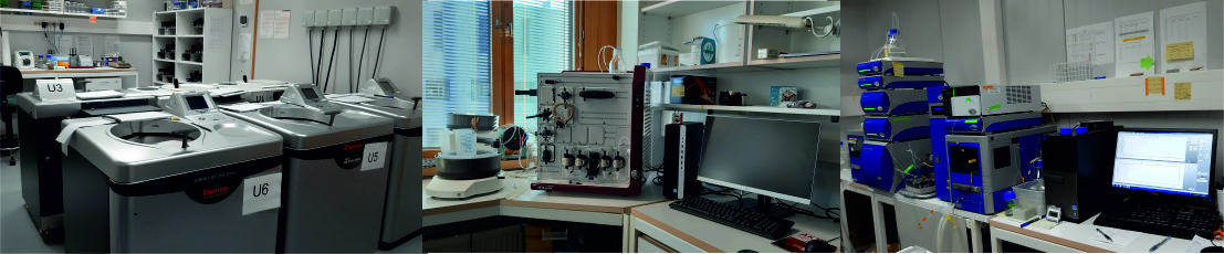
The platform also provide access to gene tagging with the CRISPR-Cas9 system for introduction of affinity tags to facilitate the purification of endogenous complexes or for tagging proteins with fluorescent reporters in view of imaging and functional proteomics applications, in the frame of a partnership with the TacGene platform from the Museum d’Histoire Naturelle (Paris, France),
The Membrane Protein Laboratory (MPL) at the Research Complex Harwell is a research and training user facility open to scientists from laboratories anywhere in the world that are interested in the Structural Biology of Membrane Proteins. The laboratory combines recently developed high throughput technologies for membrane protein production with sample preparation for crystallisation and cryo-electron microscopy. As a Wellcome funded support facility within Diamond Light Source we are uniquely placed to utilise the latest state-of-the-art developments in X-ray diffraction data collection and cryo-electron microscopy.
The Cell Free expression platform of IBS is devoted to large scale production (milligrams quantities) of soluble proteins, membrane proteins and RNAs for structural studies (X-ray, NMR, EM,...). The platform benefits from scientific input of the Membrane Transporters and Membrane and Immunity teams for expression and solubilisation of membrane proteins and from the Biomolecular NMR group for isotopic labelling of proteins and RNA. The Cell Free platform has implemented a quality management process for ISO 9001: 2015 certification, a key label and a warranty for all users of well-managed activities.
The NKI Protein Facility is highly experienced in protein production using insect and mammalian cells, and assists users with the design of expression constructs considering also solubility and domain boundaries. We collected a wide variety of expression vectors, including LIC vectors for Ligand Independent Cloning. For expression of intracellular and extracellular proteins in insect cells we have SF9 and SF21 systems. Our mammalian expression system is optimised for HEK-293T cells, but we have for instance also the glycosylation free, gnT1-deficient HEK293S variety.
Access to protein expression in bacteria (E. coli) is possible with the NKI Protein Facility, but not accessible via Instruct!
Sample preparation is not only a key issue for structural and molecular biology studies, but concerns life sciences development as a whole. Instruct centre France 1 offers state-of-the-art infrastructures for the expression and production of biomolecules and their complexes. The center proposes recombinant expression of protein and multi-protein complexes in prokaryotic and eukaryotic hosts as well as biomass production for isolation of poorly abundant proteins and endogenous complexes.
The core facility for protein production provides comprehensive services, covering every step from DNA to purified protein. These include gene cloning into expression vectors using various techniques and heterologous expression in Escherichia coli for bacterial expression, HEK293T, Expi293, and Expi293F™ GnTI- for mammalian expression, and the baculoviral system in Sf9 or High Five™ insect cells. The expression is followed by multistep protein purification.
Protein production in bacteria, baculovirus, and mammalian cells. Proteins are typically purified following a standard 3 step protocol which includes: affinity capture, ion exchange and size exclusion. Specific techniques can be employed depending on the protein, the experimental requirements, tags and yields. Removal of tags with TEV protease can be employed.
BMRZ offers extensive biochemical high-quality RNA preparation, including selective deuteration, ¹⁵N/¹³C isotope enrichment, and segmental labeling via one-step ligation of in vitro transcribed RNAs.
The HT Robotein® facility at CIP and SBBC has the particularity to combine high technology laboratory instrumentation: a central liquid handling workstation, a colony picking instrument, various microplate spectrophotometers, an Octet HTX, a LabChip GXII, a protein spotter and an Infrared Imager. This specific combination of equipment offers versatile tools for the development of numerous HT applications. Here is a list of the main services that Robotein® can provide. Nonetheless, we are always open to develop new and innovative assays.
We have developed well-established protocols for automated transformation of various bacteria and yeast strains, and for site-directed or random mutagenesis. Our platform allows automated cell cloning, high throughput colony picking, as well as cell line generation. Following automated colony picking, cultures in DeepWell plates allow selection of the best producers, together with screening for optimal culture medium. The highest expression yields can be accurately and rapidly tested on the LabChip GXII and/or the Octet HTX instruments.
Expressed proteins can be purified in a 96-well format on the robotic workstation. Automation of this process increases throughput at each purification step, reduces human error and results in higher reproducibility with an increase in yield. The efficiency of the different purification steps can be assessed on the LabChip GXII and/or the Octet HTX equipment.
Crystallographic Fragment Screening at Diamond’s XChem facility and beamline I04-1.
Supporting 1000+ fragment screens and structurally enabled fragment hit-to-lead development.
Instruct-ERIC offer a wide range of X ray approaches to determine the three dimensional shape of proteins at the atomic level.
The 'From Sample to Bio-SAXS' platform at EMBL Hamburg offers services for sample preparation for SEC-SAXS and/or automated SAXS data collection, real-time determination of physical parameter of molecules in solution and automated pipelines for building low resolution models against SAXS data.
The 'From Sample to X-ray Diffraction' platform at EMBL Hamburg offers services for obtaining crystal structures from molecules of interest. Services include: sample characterization by various biophyscial methods, sample optimization, high-throughput crystallization, robotic crystal harvesting, diffraction data collection on synchrotron beamlines, data processing, and support in structure solution and refinement.
EMBL Hamburg operates three beamlines at the PETRA III synchrotron ring, two for macromolecular X-ray crystallography and one for small angle X-ray scattering on biological macromolecules. Academic access to the beamlines is available to all research groups, and is prioritised on scientific grounds only. All proposals for beamline access are evaluated by the Project Evaluation Committee.
EMBL beamlines also offer possibilities for mail-in and remote access. Users are encouraged to contact the beamline responsibles for further details.
The Sample Preparation and Characterisation (SPC) facility supports users on the beamline and in addition offers expertise to check and improve the samples that are submitted to the crystallisation facility and the SAXS beamline.
P11 is a 3rd generation synchrotron beamline for High-throughput Macromolecular Crystallography (MX). It is energy tunable in the range 5.5-26 keV and has adaptable optics for focused beam size varying from 200 µm to 5 µm, with a maximum flux of 1013 photons/second. The large capacity sample changer can hold 368 samples in Unipuck format. The state-of-the-art photon counting detector, Eiger2 X 16M, also contributes to the high throughput of the beamline, at which 720 samples can be measured per day, making it ideal for large scale campaigns. Experimental phasing is supported with fluorescence detector. The capabilities of the beamline allow serial crystallography which is realised using a tapedrive as sample delivery and facilitated by auto-processing routines. In classical MX, the beamline can be operated remotely, and unattended data collections are under development. The beamline is supported by a user laboratory that includes OLTShifter for semi-automated crystal harvesting.
Beamlines XALOC and XAIRA are dedicated to macromolecular crystallography (MX) to provide the 3D structures of proteins, oligonucleotides and protein-protein, protein-DNA or protein-ligand complexes.
The two MX beamlines, XAIRA and XALOC, share resources to tailor the project needs through a joint proposal submission system. A new dewar shipment system and a new data portal to provide a single access point, automated processing and a catalog identification for all data acquired following FAIR data principles are being implemented to improve user experience.
EMBL Grenoble offers access to a fully automated protein to structure pipeline based on the CrystalDirect technology an the CRIMS software. Starting from purified samples, this pipeline integrates automated crystallization screening, crystal growth optimisation, automated crystal harvesting and cryocooling and X-ray diffraction data collection in a continuous and fully automated workflow operated over the Internet.
The ‘Sample Delivery Test for Serial Crystallography’ service provides users with the opportunity to evaluate different delivery methods and to refine their experimental strategies accordingly. In addition, facility staff offers guidance for drafting beamtime proposals, helping researchers to clearly articulate the technical aspects of sample preparation and delivery.
Sample delivery plays a crucial role in achieving successful data collection at XFELs. This service focuses on identifying and optimizing suitable delivery techniques for each sample type.
Our staff will perform sample delivery tests of users' samples to evaluate compatibility with various systems, including liquid jets, droplet generators, high-viscosity extruders, and fixed-target setups. During these tests, parameters such as flow stability, sample consumption, and jet behaviour are assessed to determine optimal experimental conditions. Facility staff offers these tests, ensuring that users can achieve reproducible and reliable sample delivery performance.

ALBA, the Spanish synchrotron facility, is fully committed to serve the worldwide scientific and industrial communities with state-of-the-art instrumentation and services to address the challenges of our time, most notably in Life Sciences. ALBA is currently operating 11 beamlines, with three more under design or construction phase. Since the start of user operation in 2012, ALBA has catered 8,500 researchers, half of them from European and international institutes. Three beamlines, MISTRAL, XALOC and XAIRA, are of special relevance for the activities of the Instruct-ERIC initiative.
Diamond provides a range of techniques for academic and industrial researchers studying the machines of life. As one of those techniques, Macromolecular Crystallography (MX) reveals the shape and arrangement of biological molecules at atomic resolution, knowledge of which provides a highly accurate insight into function. This can be combined with complementary information from many other techniques available at Diamond alongside lab based investigations to reveal the broader picture of molecular interactions and their effects..
Small Angle X-ray Scattering (SAXS) covers the major disciplines of Biology, Chemistry and Physics delivering structural and dynamic information in nanoscience, mesoscopic architectures, supramolecular structures and nucleation/growth of crystals. SAXS is also proving important in archaeological, environmental and conservation sciences, indicating an ability to span a wide range of scientific disciplines.
The 'X-ray Diffraction and Bio-SAXS ' platform at EMBL Hamburg offers beamtime on one beamline for small-angle X-ray scattering (P12) and two beamlines for macromolecular crystallography (P13, P14). With both methods, static and time-resolved pump-probe experiments can be performed.
Diffraction techniques core facility provides services for characterization of protein structure using single crystal X-ray diffraction and small-angle X ray scattering for analysis of liquid samples. Both methodes are backer by advanced approaches available in laboratory. The quality of high-intense X-ray beams is ensured by using of MetalJet sources (Excillum). The provided services include assistance with data processing and reporting ready for publication.
Regular access to synchrotron beam time for MX experiments (remote, on-site, mail-in)
Crystallographic fragment screening, either to-go or on-site
Biocenter Oulu Protein Crystallography core facility offers home-source X-ray generator for macromolecular crystal diffraction experiments.
Crystallographic Fragment Screening at Diamond’s XChem facility and beamline I04-1.
Supporting 1000+ fragment screens and structurally enabled fragment hit-to-lead development.